Degenerative and isthmic spondylolisthesis share a name and a picture: one vertebra sits forward of the one below it. They do not share a cause, a typical age, or the same crowded nerve space. Treating them as one condition is how the wrong explanation gets attached to a slip.
Grades, the full list of causes, and the general workup are in What Is Spondylolisthesis? This article is only the difference between the two types adults actually have.
Two mechanisms, not two sizes
The Wiltse system, published in 1976, sorts slips by cause. Isthmic means the pars interarticularis, a thin bridge of bone at the back of the vertebra, is cracked or lengthened. Degenerative means that bridge is intact and the disc and facet joints have worn enough to let the vertebra creep forward.
One is a broken hook. The other is a worn joint. The percentage of slip, the Meyerding grade, is a separate measurement and does not tell you which mechanism you have.
That difference decides what moves. When the pars is intact, the back of the vertebra, including the lamina and the facets, slides forward with the body. The canal in front of that bone gets smaller.
When the pars is broken, the body can slip forward while part of the back stays behind. The central canal may be less the issue, and the opening where a nerve root leaves may be more the issue. StatPearls describes these as distinct types. A report that never says which one you have has not finished the diagnosis.
At a glance
Degenerative and isthmic slips are different problems
The names describe the cause, not the size of the slip. Grade measures distance. Neither name is an order for surgery.
| Question | DegenerativeWorn disc and facets | IsthmicA crack in the bridge |
|---|---|---|
| The bone bridge | Intact. Worn disc and facets allow the slip. | Cracked or stretched. A defect does not always produce a slip. |
| Who is usually affected | Older adults. | Often begins in adolescence. |
| Usual level | Often L4-L5. | Often L5-S1. |
| Where the nerve is crowded | The canal often narrows because the back of the vertebra moves forward too. | The nerve's exit is the space to look at. |
Grade measures distance. Type names the cause. Neither one is a surgical order.
Who has which, and at which level
Degenerative slips show up as the spine ages. In a Framingham CT study of 188 adults aged 40 to 80, degenerative spondylolisthesis was about three times more common in women than in men, and prevalence rose from the fifth through the eighth decades. StatPearls notes that this type most often sits at L4-L5. A slip at L4-L5 in a postmenopausal woman is not proof of the degenerative type, but it is the pattern to expect until the pars is actually inspected.
Isthmic slips often begin much earlier. Repeated extension in sport, gymnastics, football, wrestling, can stress the pars in adolescence. Many defects stay silent for years and are found on an adult scan done for something else.
In that same Framingham analysis, spondylolysis was present in 11.5% of adults, and about a fifth of people with defects on both sides had no measurable slip. The defect and the slip are not the same finding. Isthmic slips, when they do occur, favor L5-S1 and are more often seen in men.
Level is a clue, not a diagnosis. L4-L5 can be isthmic. L5-S1 can be degenerative.
The images have to show the pars. If your report says “spondylolisthesis at L4-L5” and stops, ask whether the pars is intact.
Why the nerve symptoms differ
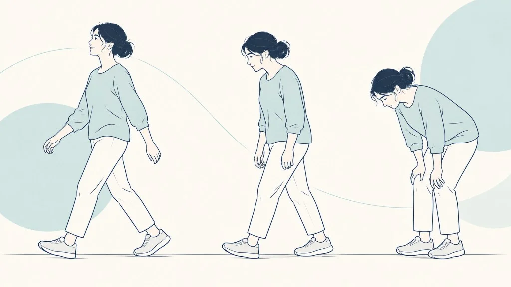
People with degenerative slips often describe the walking pattern of lumbar stenosis: leg heaviness that builds with walking and eases with sitting. NIAMS describes that pattern. The slip is one reason the canal is tight, along with thickened ligament and swollen joints. How those two diagnoses travel together is the subject of Spondylolisthesis and Spinal Stenosis Together.
People with isthmic slips more often have one-sided leg pain in the root that exits at that level, or back pain tied to extension, or no pain at all. A teenager with activity-related back pain and a pars reaction on MRI is at the start of this path. An adult with an old defect and no leg symptoms is further along a path that may never need treatment. Do not import the stenosis story into an isthmic slip just because both reports contain the word spondylolisthesis.
| Question | More often degenerative | More often isthmic |
|---|---|---|
| Bone bridge (pars) | Intact | Cracked, lengthened, or a stress injury |
| Typical level | L4-L5 | L5-S1 |
| Typical age | Older adults | Defect often starts in adolescence; slip may be found later |
| Nerve space | Central canal, with a stenosis pattern | The exit opening of a nerve root |
| What it does not decide | Whether you need surgery, a fusion, or any treatment aimed at a symptom you do not have | |
What the type should change, and what it should not
It should change the scan review. Look at the pars. Look at the canal if the pars is intact, and at the foramen if it is not. It should change the history you emphasize: walking tolerance in a degenerative slip, sport and extension in a young isthmic spine.
It should not change a stable, tolerable life into an operation. A grade 1 slip of either type can be observed when symptoms are mild and strength is stable. What “grade 1” does and does not require is covered in Grade 1 Spondylolisthesis: Does It Need Surgery? It should also not pick fusion by reflex.
Fusion is a proposal about motion and about how much bone a decompression would remove. The type informs that proposal. Your goals and the exam decide it.
If you are preparing for a visit, the single most useful question is the type. The questions to prepare help you ask it without treating the answer as a candidacy score. Write down whether anyone has said “pars intact” or “pars defect.” Those four words sort the rest of the conversation.
How the visit should sound different
A degenerative-slip visit spends its time on walking minutes, whether a cart helps, and whether the canal on MRI matches those minutes. The slip grade is mentioned, then set beside the stenosis. Bending films matter if someone is about to discuss holding the level still.
Hip motion and foot pulses belong in the same hour, because both imitate a walking limit. You should leave knowing whether the pars was seen and called intact.
An isthmic visit spends its time on the pars itself. In a teenager, that means how long the pain has tracked the sport, whether MRI shows a stress reaction or a completed gap, and what a break from extension looks like for this season. In an adult, it means whether an old defect is actually the cause of today’s pain or an incidental scar.
One-sided leg pain in a root pattern pulls the eye to the foramen. A painless defect does not become a training ban.
If the visit never separates those scripts, ask for the separation. “Which type, and what does that change?” is a fair question. An answer that only restates the percent has not used the type.
The percent is shared vocabulary. The mechanism is the decision.
What you can ignore until the type is known
Ignore exercise lists that do not say which slip they were written for. Extension drills that are reasonable for one back are the load that irritates a pars. Flexion-biased walking advice that helps stenosis can be irrelevant to a quiet isthmic defect.
Ignore surgical slogans too. “Slips need fusion” and “slips never need fusion” both skip the pars. The studies people quote about fusion and decompression were largely done in degenerative slips with stenosis, not in adolescent pars injuries. Importing those results into a teenager’s X-ray is a category error.
Ignore, also, the fear that the type is a permanent identity. A pars defect does not progress to a degenerative slip by changing its name. They are different structures.
You can have ordinary age-related discs above an old isthmic level. You can have a degenerative slip and a disc bulge at the next level that has nothing to do with today’s leg. The type tells you which finding deserves the first explanation. It does not freeze the rest of the spine in place.
Bring the images, not only the impression line. The impression is where “spondylolisthesis” survives and “pars intact” disappears. Open the axial pictures with the clinician and ask to see the bridge of bone. Four words, seen rather than remembered, are worth more than a second opinion that only repeats the grade.
A second opinion is most useful when surgery is being offered or when two clinicians have named two different types. It is less useful as a search for a gentler grade. Bring the same standing film and the same MRI.
Ask both people to say the type, the tight space, and the symptom they think they are treating. If those three match, you have a coherent story even if the adjectives differ. If they do not match, you do not yet have a plan.
You have two readings. Stay with the reading that can point at the pars and at your walking notes in the same sentence. That is the whole difference between these slips, applied to one person.
Sex, age, and level are clues with overlap, so do not let them substitute for the images. The Framingham CT analysis found degenerative slips about three times as often in women, rising with age, and found pars defects in 11.5 percent of the adults scanned. StatPearls places the typical degenerative level at L4-L5.
Isthmic slips more often sit at L5-S1. A man in his seventies can still have an old pars defect. A woman in her forties can still have a degenerative slip.
If a report guesses the type from the demographic and never mentions the pars, ask for the pars. Demographics are how these slips were described in populations. They are not how your film is read.
Once the type is secure, stop re-deriving it at every appointment. Put it in the first sentence you tell the next clinician, along with the grade and the symptom. “Grade 1 degenerative slip at L4-L5, pars intact, legs heavy after ten minutes” is a usable opening.
“Grade 1 isthmic at L5-S1, old pars defect, back pain only with extension, no leg symptoms” is a different opening. Both are spondylolisthesis. They should never again be allowed to share a treatment paragraph just because they share a noun.
If a future note drops the type, put it back in the margin yourself. The next reader of that note may be you, a year from now, trying to remember which problem you actually have. Keep the type where you can find it. A year from now, that one word will save you from starting the explanation over.
Frequently asked questions
Which type is more serious?
Neither label is more serious on its own. A small degenerative slip that crowds the canal and stops you walking can limit life more than a long-standing isthmic slip that is quiet. A high-grade isthmic slip in a teenager is a different conversation from a grade 1 degenerative slip in a 70-year-old. Seriousness is symptoms, nerve findings, and whether the level moves, not the Wiltse type printed on the report. Ask which type you have so the rest of the plan matches the cause.
Can imaging tell degenerative from isthmic?
Usually yes. A defect in the pars, the small bridge at the back of the vertebra, points to isthmic. An intact bridge with worn facets and a slip points to degenerative. Standing side-view X-rays show the slip. CT shows the bone defect most clearly. MRI shows the nerves and the discs. A report that says spondylolisthesis without saying which type is incomplete. The two types are not treated as interchangeable once the images are read carefully.
Do both types cause spinal stenosis?
They can both crowd a nerve, in different places. In a degenerative slip the pars is intact, so the back of the vertebra moves forward with the body and the central canal often narrows. That is why degenerative slips and lumbar stenosis so often arrive together. An isthmic slip, with a broken bridge, more often affects the opening where a nerve root leaves. Either pattern can hurt. Neither pattern means you need an operation today.
I was told I have a pars defect. Is that the same as a slip?
No. A pars defect is spondylolysis, a crack or stress injury in that bridge of bone. Spondylolisthesis is the slip that sometimes follows. In a Framingham CT study, the prevalence of spondylolysis was 11.5%, and about a fifth of people with defects on both sides had no measurable slip. A defect without a slip, and without symptoms, is a finding. It is not a smaller version of the degenerative slip seen in older adults.
Does the type decide between decompression and fusion?
It narrows the question. It does not answer it. Degenerative slips are discussed alongside stenosis, and trials in that group disagree about when fusion adds benefit. Isthmic slips raise a different stability question because the bone bridge is already broken. Grade, motion on bending X-rays, which nerve space is tight, and your symptoms still have to be added. A sentence that says 'isthmic, therefore fusion' or 'degenerative, therefore no fusion' is too short.
Key takeaways
- Degenerative slips are usually later in life and at L4-L5. Isthmic slips usually start with a pars defect and favor L5-S1.
- An intact pars tends to narrow the central canal. A broken pars more often affects a nerve's exit.
- Many pars defects never become a slip, and many slips never become a surgery.
- The type is the first sorting step. Symptoms, motion, and the nerve exam still decide treatment.
References
- Margetis K, Gillis CC. Spondylolisthesis. StatPearls, National Library of Medicine (NCBI Bookshelf).
- Wiltse LL, Newman PH, Macnab I. Classification of spondylolisis and spondylolisthesis. Clinical Orthopaedics and Related Research. 1976;(117):23-29. 1976.
- Kalichman L, Kim DH, Li L, Guermazi A, Berkin V, Hunter DJ. Spondylolysis and spondylolisthesis: prevalence and association with low back pain in the adult community-based population. Spine. 2009;34(2):199-205. 2009.
- Spinal Stenosis. National Institute of Arthritis and Musculoskeletal and Skin Diseases (NIAMS), National Institutes of Health.
Prepare questions for your consultation
Organize your symptoms, imaging, and treatment history into clear questions for a spine specialist. The questionnaire does not decide candidacy.