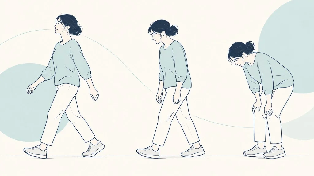
A narrow lumbar canal on an MRI is easy to see and easy to over-read. The diagnosis of lumbar spinal stenosis is the harder step: deciding that the narrowing explains this person’s walking limit. Clinicians do that by putting the history and the examination next to the pictures. Any one of the three, alone, is not enough.
This article stays on that pathway. The condition itself, including what the canal is and how common narrowing is, is explained in What Is Lumbar Spinal Stenosis?
The history that makes the diagnosis plausible
The story clinicians listen for is specific. Leg pain, heaviness, or numbness builds while you walk or stand. It eases when you sit, or when you lean forward on a cart.
You may prefer walking slightly bent. Back pain can be present, but back pain that never changes with walking is a weaker fit. NIAMS describes this pattern, and a JAMA review treats a history of back and leg pain provoked by extension and relieved by flexion as the usual starting point, with imaging used to support it.
Details that sharpen the story are the distance you can walk, whether a shopping cart helps, whether hills are easier downhill than up, and whether the limit is getting shorter. A limit measured in minutes is more useful than the word “severe.” How those severity words differ from function is covered in Mild vs. Moderate vs. Severe Lumbar Spinal Stenosis.
The history also looks for other explanations. A groin ache and a limp when the hip is rotated point toward the joint, discussed in Is Your Lower Back Pain Coming From Your Spine or Your Hip? A calf cramp that stops when you stand still, rather than when you sit, raises the possibility of poor arterial flow. Night pain, unexplained weight loss, fever, or a history of cancer are reasons to look beyond ordinary wear.
Process
Each step checks the one before it
Walking or standing that brings on leg symptoms, eased by sitting or leaning forward, is what makes the diagnosis plausible. Imaging comes after that story, and only when it will change a decision.
- Symptoms and history. Walking or standing brings on leg symptoms. Sitting or leaning forward eases them. Back pain alone is not the syndrome.
- Physical and neurological examination. Strength, reflexes, sensation, hip motion, and foot pulses. Weakness changes urgency. A normal strength exam does not erase a clear history.
- Imaging when it will change a decision. Usually MRI, to see whether a canal or a nerve opening is narrow at a level that could explain the legs.
- Clinical correlation. The narrow level has to fit the story and the exam. A crowded canal in a comfortable walker is a finding, not a diagnosis.
- Treatment discussion. Only after the match. The diagnosis does not by itself mean an operation, an injection, or a device.
What the examination adds
The exam looks for signs the history cannot confirm. Strength at the ankle and the big toe, the knee reflex and the ankle reflex, and sensation on the foot can show that a particular nerve root is involved. A weak foot that slaps the ground is more urgent than pain of the same intensity with full strength.
Many people with symptomatic stenosis still have a normal strength exam on a given day. The walking history can carry the diagnosis when the exam is quiet.
The same visit should move the hip and feel the pulses. Hip arthritis and blocked leg arteries both limit walking, and both are common in the same age group as stenosis. AAOS OrthoInfo notes that the evaluation includes the history, the exam, and imaging, and that other causes of leg pain are part of sorting this out. If pulses are diminished, a vascular opinion belongs in the workup before a spine procedure is planned.
Bowel or bladder changes, numbness in the saddle area, or rapidly worsening weakness are not routine stenosis. They need same-day care. They are uncommon. Mentioning them is part of a careful history, not a prediction that they will occur.
What MRI is for, and what it is not
MRI shows the canal, the side openings, the discs, and the nerves without radiation. It is the usual study when a procedure is being considered or the diagnosis is uncertain. CT is used when MRI is unsafe or unavailable, and X-rays show alignment, including any slip of one vertebra on another, which changes the conversation if surgery is later discussed.
The scan answers a narrow question: is there a place where the nerves have less room, and does that place match the legs? It does not grade your disability. A Framingham CT study found relative narrowing in 22.5% of community adults and absolute narrowing in 7.3%, rising sharply after age 60, with many people reporting no symptoms. That is why a report that says stenosis, handed to you before anyone has asked how far you walk, is incomplete.
The words mild, moderate, and severe on that report are a separate problem of language. They should not be treated as the diagnosis getting worse or better. Correlation is the sentence a clinician should be able to say out loud: this level is narrow, this is where your symptoms are, and this is what we are hoping to change.
| Step | What it can show | What it cannot show |
|---|---|---|
| Walking history | Whether posture and distance provoke and relieve the legs | Which level is narrow, or whether an artery or a hip is the limit |
| Neurological exam | Weakness, reflex change, or a sensory pattern, and how urgent it is | The cause, when the exam is normal, which is common |
| Hip and pulse check | A joint or a circulation problem that can mimic the walk | Whether the canal is also narrow. Both can be true |
| MRI or CT | Where the canal or a nerve opening is tight | That the tightness is why you hurt, or which treatment you need |
Tests that sometimes get added, and why
Electrodiagnostic studies, nerve conduction and needle examination, are not routine for a classic walking history and a matching MRI. They become useful when the story could be a peripheral nerve instead, when diabetes or another neuropathy is in the background, or when the exam and the images disagree. A normal study does not erase stenosis.
An abnormal one does not choose the operation. It is a tie-breaker, and only sometimes.
Flexion and extension X-rays are a different add-on. They look for a slip that moves, which matters if someone is discussing surgery, because a moving slip raises the question of stability. They are not required to recognize the walking syndrome. Ordering them “just to be complete” before any surgical conversation adds radiation without a decision to inform.
Blood tests do not diagnose stenosis. They are for the other list: infection, inflammatory disease, or problems that can mimic back and leg pain. A clinician who orders them is usually chasing a feature of your history, such as fever, morning stiffness, or weight loss, not confirming a narrow canal.
If no one has explained why a test was ordered, ask. The answer should name the alternative they are trying to exclude.
What to gather before the visit
The most useful record is a week of walking notes. How many minutes to the first leg symptom, whether a cart or a lean helped, and whether you recovered by sitting. One number, repeated, beats a long adjective.
Add a list of medicines, any hip or vascular history, and the actual imaging disc or portal link. A printed impression without the images forces the next clinician to repeat the scan or to trust a word they cannot see.
Write down what you want the visit to decide. Some people need a name for the walking limit. Some need to know whether the hip should be examined first.
Some already know the name and want to know whether an injection or an operation is even on the table. Those are different appointments. Arriving with “my report says severe” as the only question tends to produce a long explanation of an adjective and a short discussion of your week.
After the match is made
A diagnosis of symptomatic stenosis opens a treatment conversation. It does not close one. Many people start with activity changes and therapy.
Some later consider an injection or a decompression. A few, if a vertebra is slipping or an operation would leave the segment unstable, hear about fusion. None of those steps is implied by the MRI sentence alone.
If you are preparing for that conversation, the useful materials are the walking distance, a list of what eases it, the actual images, and any hip or vascular history. The questions to prepare are a way to organize that. They are not a score that says you do or do not have stenosis.
A diagnosis you can repeat in one sentence
Leave the visit able to say one sentence a relative would understand. “My legs ache after about ten minutes of walking, sitting fixes it, and the MRI shows a tight canal at a level that fits.” Or: “The canal is tight, but my limit is actually the hip, and the spine is not the first target.”
Or: “We are not sure yet, because the pulses are hard to feel and a vascular clinic is next.” Each of those is a diagnosis in use. “I have severe stenosis” is a fragment of a report.
If the sentence has a blank where the walking time should be, you do not have a baseline. Fill it in over the next week and bring it back. If the sentence names a level the exam never tested, ask for the strength and reflex check before anyone discusses a procedure at that level.
The order is history, exam, images, match. Skipping to the match because the report already used the word stenosis is how the wrong level, or the wrong joint, gets treated.
Second opinions are reasonable when surgery is on the table or when two clinicians describe different sources for the same walk. They are less useful as a hunt for a milder adjective. Bring the same images to both visits so the disagreement, if there is one, is about interpretation rather than about two different scans.
Write down what each person thinks is causing the limit and what they expect a treatment to change. Agreement on those two points matters more than agreement on mild versus moderate.
If the sentence you leave with still depends on a word you do not understand, stop and ask for a translation before you agree to the next test. “Severe” should be restated as which space is tight and how that fits the minutes you can walk. A translated diagnosis is one you can use.
An untranslated one only feels like news. The overview of what stenosis is, if you still need it, stays in the companion article. This page was only the path that gets you to a sentence you can trust. Where the narrowing sits, in the center of the canal or in the side recess, is a separate description, covered in Central vs. Lateral Recess Spinal Stenosis: What’s the Difference?.
Frequently asked questions
Can lumbar stenosis be diagnosed without an MRI?
The clinical pattern can be recognized from the history and the exam, and a review in JAMA describes the diagnosis as generally made that way, then supported by imaging. Treatment decisions that involve an injection or an operation need pictures, usually MRI, so the narrow level can be identified. X-rays show alignment and bone, not the canal well. CT is an alternative when MRI is not possible. A scan is not required to start walking advice or physical therapy when the story is typical and there are no urgent nerve findings.
What if my MRI shows stenosis but my legs feel fine?
Then you have a radiographic finding, not the clinical syndrome. Community CT studies have found narrowing in a substantial share of older adults, including people without symptoms. Treating a picture in a person who walks comfortably exposes them to the risks of treatment without a symptom to improve. Keep the report. Seek care if a walking limit, leg numbness, or weakness appears. Do not schedule a procedure to 'fix' a canal that is not limiting you.
How do doctors tell stenosis from poor circulation?
Both can limit walking. Stenosis symptoms often ease within a short time of sitting or leaning forward, and pulses in the feet are usually present. Vascular claudication is more often a cramp in the calf that eases when you simply stop, even if you stay standing, and foot pulses may be weak. The two can coexist. If the pulses are hard to feel, or the pain is a calf cramp that ignores posture, a vascular evaluation belongs in the workup before the spine is treated.
Does a normal neurological exam rule stenosis out?
No. Many people with symptomatic stenosis have pain and a walking limit with a normal strength exam on the day they are seen. Weakness, a missing reflex, or numbness in a root pattern supports a nerve problem and can change urgency. Their absence does not erase a clear walking history that matches a narrow canal. The exam is one part of the match, not a pass-fail test.
Should I get an MRI before I see a clinician?
It is usually more useful the other way around. A scan ordered without a history often finds age-related narrowing that then has to be explained, and it can miss the question that actually needed answering, such as the hip or the arteries. If you already have a recent MRI, bring it. If you do not, a clinician can decide whether pictures will change the next step. Urgent symptoms, including new bowel or bladder trouble or rapidly worsening weakness, should not wait on a routine scan appointment.
Key takeaways
- The diagnosis is a match: walking history, examination, and imaging that fits.
- MRI shows the canal. It does not, by itself, explain the symptoms or choose the treatment.
- A normal strength exam does not rule the syndrome out. Urgent nerve changes raise the pace.
- Hip disease and poor leg circulation can limit walking and need to be considered.
References
- Katz JN, Zimmerman ZE, Mass H, Makhni MC. Diagnosis and Management of Lumbar Spinal Stenosis: A Review. JAMA. 2022;327(17):1688-1699. 2022.
- Spinal Stenosis. National Institute of Arthritis and Musculoskeletal and Skin Diseases (NIAMS), National Institutes of Health.
- Kalichman L, Cole R, Kim DH, et al. Spinal stenosis prevalence and association with symptoms: the Framingham Study. The Spine Journal. 2009;9(7):545-550. 2009.
- Lumbar Spinal Stenosis. American Academy of Orthopaedic Surgeons, OrthoInfo.
Prepare questions for your consultation
Organize your symptoms, imaging, and treatment history into clear questions for a spine specialist. The questionnaire does not decide candidacy.