Lumbar spinal stenosis is one of the most common reasons older adults are referred to a spine specialist. It is also one of the most misunderstood diagnoses, partly because the word “stenosis” appears on so many MRI reports. This overview explains what the condition is, why it develops, how it feels, how clinicians confirm it, and the broad treatment paths people consider.
What narrows the lumbar spinal canal
The lumbar spine is made up of five vertebrae stacked above the pelvis. Together they form a protective channel, the spinal canal, for the nerves that travel to the legs, bladder, and bowel. In the lower back, the spinal cord has already ended, so the canal carries a bundle of nerve roots called the cauda equina.
Stenosis means narrowing. NIH describes three places where this can happen: the central spinal canal, the space where nerve roots branch off, and the openings between vertebrae where nerves leave the spine.
In most people, narrowing develops slowly over years from several age-related changes happening together:
- Disc changes. Discs lose water and height and may bulge backward into the canal.
- Facet joint arthritis. The small joints at the back of the spine enlarge and can form bone spurs, also called osteophytes.
- Ligament thickening. The ligamentum flavum, a ligament lining the back of the canal, can thicken and buckle inward.
- Vertebral slippage. A vertebra can slip forward on the one below, a condition called spondylolisthesis. It frequently occurs alongside stenosis and is explained in What Is Spondylolisthesis?
Less commonly, stenosis results from a naturally narrow canal present from birth, previous injury or surgery, or conditions such as Paget’s disease or rheumatoid arthritis.
Where the narrowing occurs
Clinicians often describe stenosis by location, because location influences symptoms and surgical planning.
- Central stenosis affects the main canal and can cause symptoms in both legs.
- Lateral recess stenosis affects the side channel where a nerve root travels just before it exits, and often causes symptoms on one side.
- Foraminal stenosis affects the opening where a nerve root leaves the spine.
One person can have more than one type, and narrowing can affect more than one level of the lower spine.
How common it is, and why imaging alone can mislead
Narrowing on imaging is common, especially with age, and it does not always cause symptoms.
In a Framingham Study analysis of CT scans from 191 community participants, acquired narrowing that met a “relative” stenosis threshold was found in 22.5% and a stricter “absolute” threshold in 7.3%. Among participants aged 60 to 69, those figures were 47.2% and 19.4%. Absolute stenosis was associated with low back pain, but many people with narrowing on CT did not report symptoms.
The clinical condition, meaning narrowing plus typical symptoms, is less common than the imaging finding. A 2022 review in JAMA estimates that the clinical syndrome affects approximately 11% of U.S. adults and becomes more common with age.
The practical point is simple. Stenosis on an MRI report is a description of anatomy. Whether it explains your symptoms depends on whether the location and pattern fit.
Symptoms: what lumbar spinal stenosis feels like
Symptoms usually develop gradually. Some people with narrowing have no symptoms at all.
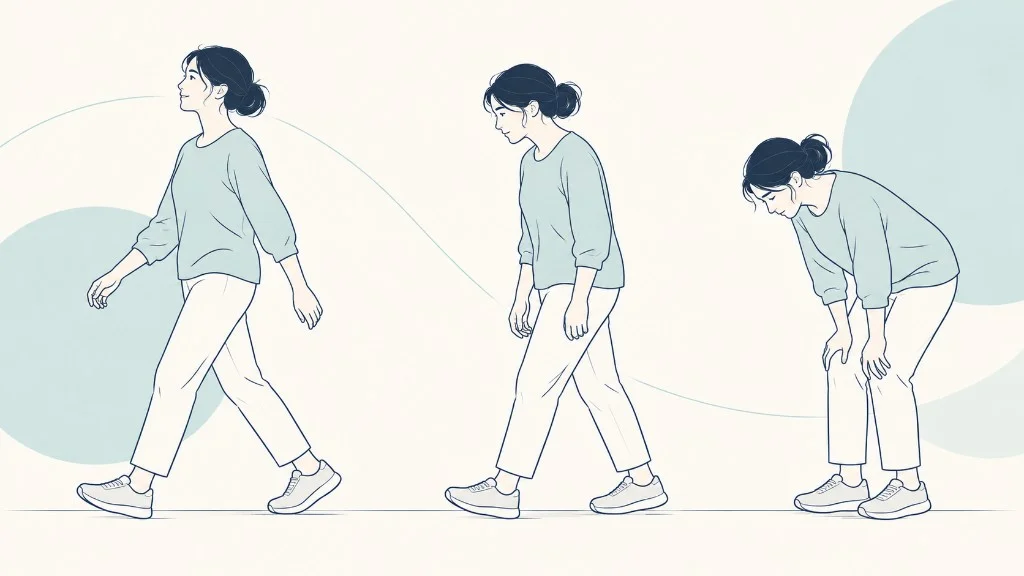
The hallmark symptom is neurogenic claudication, which is leg pain, heaviness, numbness, or weakness caused by pressure on nerves in the lower spine. It typically:
- starts in the buttocks and spreads into the thighs or lower legs
- builds with standing or walking, especially walking upright
- eases after sitting or bending forward for several minutes
- affects one or both legs
Many people find they can walk farther leaning on a shopping cart or ride a stationary bike comfortably. NIH explains that bending forward opens up the spinal column and enlarges the spaces at the back of the spine, which is why these positions help.
Symptom pattern
What neurogenic claudication usually does
The hallmark is leg pain, heaviness, numbness, or weakness from pressure on the nerves. Position changes it. Narrowing on a scan does not, by itself, mean this pattern is present.
What brings it on
- Standing or walking, especially upright
- Often starts in the buttocks and spreads down the leg
- One leg or both
What eases it
- Sitting or bending forward for several minutes
- Leaning on a shopping cart
- A stationary bike is often comfortable
What may be missing
- Some people with narrowing have no symptoms
- Low back pain can occur, but it is not the hallmark
- A scan word is not the same as a walking limit
Bending forward opens the spaces at the back of the spine, which is why those positions help.
Other possible symptoms include low back pain, tingling or cramping in the legs and feet, and leg weakness. Position-dependent back pain has several possible causes besides stenosis, which we compare in Lower Back Pain When Standing vs. Sitting.
Symptoms that need urgent assessment
- New loss of bladder or bowel control, or difficulty emptying the bladder
- Numbness in the groin, genitals, or inner thighs
- Leg weakness that is severe or rapidly getting worse
These can indicate severe compression of the nerve bundle, known as cauda equina syndrome. It is uncommon but is treated as an emergency.
How lumbar spinal stenosis is diagnosed
Diagnosis combines three things: your history, a physical examination, and imaging.
Process
How the pieces are put together
Diagnosis combines the history, the examination, and imaging. The narrow level has to fit the person. The radiology wording does not make the decision by itself.
- HistoryWhere symptoms are felt, what brings them on, and what eases them, including walking distance.
- ExaminationStrength, reflexes, sensation, and walking, plus other causes such as the hip or circulation.
- Imaging when it will change decisionsUsually MRI. Narrowing is common in people without symptoms, so the picture is matched to the person.
- A treatment conversationBased on how limiting the symptoms are, not on the radiology wording alone.
History. A clinician will ask where the pain is, where it travels, how far you can walk, what positions help, and whether you have numbness, weakness, or bladder changes. The JAMA review notes that the diagnosis can generally be made from a history of back and leg pain provoked by extension and relieved by flexion, then confirmed with cross-sectional imaging.
Examination. This usually includes strength, sensation, and reflex testing in the legs, walking observation, hip examination, and checking foot pulses. Pulses matter because narrowed leg arteries can cause walking-related leg pain that resembles stenosis.
Imaging. MRI is the most common test because it shows discs, ligaments, and nerves. CT, or CT myelography when MRI is not possible, is an alternative. Standing or bending X-rays can show slippage or abnormal movement between vertebrae.
Imaging is usually most useful when symptoms persist, when a procedure is being considered, or when a serious cause needs to be excluded. The report may grade narrowing as mild, moderate, or severe. Those grades describe the picture, not how much you are affected, and they are not a treatment recommendation on their own.
How stenosis tends to change over time
For many people, lumbar spinal stenosis is not a steadily worsening condition. The JAMA review summarizes a series of patients followed for up to three years without surgery: approximately one-third reported improvement, about half reported no change, and roughly 10% to 20% reported that their back pain, leg pain, and walking were worse.
Because change is usually gradual, most people have time to try non-surgical treatment and reassess. The exceptions are the urgent symptoms listed above and weakness that is clearly progressing.
Treatment options and their trade-offs
Treatment is guided by how much symptoms limit daily life, how they respond to initial care, the imaging findings, and personal goals. The table below summarizes the main approaches.
| Approach | Goal | Potential benefit | Important limitation |
|---|---|---|---|
| Activity modification, medications, physical therapy | Reduce symptoms and maintain function | First-line care with low risk; many people improve or stay stable | Does not change the narrowing itself; benefit varies |
| Epidural steroid injection | Reduce nerve inflammation and pain | May give short-term relief for some people | Long-term benefit has not been demonstrated |
| Decompression surgery (such as laminectomy) | Remove tissue that compresses the nerves | In a major trial of surgical candidates, surgery improved some outcomes more than non-surgical care; results depended on the analysis used | Surgical risks; not every symptom resolves, and some people need further surgery |
| Decompression with fusion | Relieve compression and stop motion at an unstable level | Addresses instability or deformity when present | Longer surgery, more risk and cost; stenosis alone does not make fusion necessary, and added benefit depends on factors such as instability |
| Decompression with a motion-preserving implant | Relieve compression and stabilize while keeping some motion | An option for a narrowly defined group under FDA approval | Applies only to patients who meet the labeled indication; long-term data are still accumulating |
Non-surgical care
According to the JAMA review, first-line treatment includes activity modification such as shortening periods of standing or walking, pain medications such as NSAIDs, and physical therapy. The same review states that long-term benefits of epidural steroid injections for stenosis have not been demonstrated.
The American Academy of Orthopaedic Surgeons (AAOS) adds practical detail about these options:
- Physical therapy usually combines stretching, core strengthening, and conditioning. Some people find exercise in a bent-forward position, such as riding a stationary bike, easier to tolerate than upright walking.
- Anti-inflammatory medicines can ease pain but need to be used carefully, because they can cause stomach irritation, ulcers, and kidney problems. Tell your clinician if you develop reflux or stomach pain while taking them.
- Epidural steroid injections may reduce pain and numbness, but not weakness. AAOS advises no more than three injections per year.
- Lumbar traction has very limited results, and there is no scientific evidence of its effectiveness.
- Acupuncture and chiropractic care may help some pain in less severe cases. Long-term benefit from acupuncture has not been proven, and spinal manipulation needs caution in people with osteoporosis or a herniated disc.
AAOS also points out that these treatments do not widen the spinal canal. Their purpose is to relieve symptoms and help you stay active while you and your clinician see how the condition behaves over time.
Preparing for a specialist visit
A short record kept for a week or two can make an appointment more productive. Useful details include how far or how long you can walk before symptoms start, what makes them ease, which leg is affected, any numbness or weakness, and which treatments you have already tried and for how long. How that distance is used, and why it is not a grade of stenosis, is covered in Walking Distance and Lumbar Spinal Stenosis: A Limit, Not a Grade.
What an epidural injection can and cannot change is covered in Epidural Injections for Lumbar Spinal Stenosis: What They Can Change. Bring copies or a list of previous imaging, and note any change in bladder or bowel control, even if it seems minor.
Questions worth asking include: Do my symptoms match what my imaging shows? What is the goal of the next treatment step, and how will we know whether it is working? Which symptoms would make you want to see me sooner?
Surgery
Surgery is generally considered for people whose symptoms persist and meaningfully limit daily life despite non-surgical care. The most studied operation is decompression, which removes bone and thickened tissue pressing on the nerves. What Is a Laminectomy? explains the procedure in detail.
The evidence has nuances. In the SPORT trial, which studied surgical candidates with stenosis but without spondylolisthesis, many patients crossed over between groups. The intention-to-treat analysis showed a significant benefit for surgery on bodily pain at two years, but not on physical function or the Oswestry Disability Index. The as-treated analysis, which combined randomized and observational patients and adjusted for confounders, showed significantly greater improvement with surgery on all primary outcomes.
When a vertebra has slipped and the segment is unstable, or the planned decompression could leave it unstable, surgeons may consider adding fusion to stabilize the level. A slip seen on imaging does not by itself mean fusion is needed. Another option, available for a defined group, is a motion-preserving implant placed after decompression.
The FDA approved the TOPS System (the link goes to the manufacturer’s website) in 2023 for patients aged 35 to 80 with Grade I degenerative spondylolisthesis at one level from L3 to L5 and moderate to severe stenosis, among other criteria. FDA approval defines who was studied and who the device is labeled for; it does not mean the device is appropriate for any particular person. Our clinical evidence page explains that study, its results, and its limits.
Which option makes sense depends on diagnosis, instability, anatomy, the number of levels involved, bone quality, general health, previous surgery, and what you want to be able to do. Those are questions for a spine specialist who has examined you and reviewed your imaging.
Frequently asked questions
Is lumbar spinal stenosis the same as arthritis of the spine?
They are related but not the same. Arthritis of the spine, meaning wear in the discs and facet joints, is the most common cause of lumbar spinal stenosis. Stenosis describes the result: narrowing of the spaces that hold the nerves. Many people have spinal arthritis without meaningful narrowing, and some have narrowing on imaging without symptoms. The diagnosis of symptomatic stenosis depends on typical symptoms plus imaging that shows narrowing in a matching location.
Does lumbar spinal stenosis always get worse?
No. In studies that followed people without surgery, a substantial share stayed about the same or improved over several years, while a smaller group worsened. A 2022 JAMA review summarized one such series: over up to three years, roughly a third reported improvement, about half reported no change, and roughly 10% to 20% reported worsening. Progression tends to be slow when it happens, which usually allows time to try non-surgical care and reassess.
Can lumbar spinal stenosis cause paralysis?
Lumbar stenosis affects nerve roots below the end of the spinal cord, so it does not cause the kind of paralysis associated with spinal cord injury. It can, however, cause leg weakness, and in rare cases severe compression of the nerve bundle can cause cauda equina syndrome, with loss of bladder or bowel control, saddle numbness, and leg weakness. Those symptoms need emergency assessment. Gradual weakness should also be reported promptly rather than waited out.
What does moderate or severe stenosis on my MRI report mean?
These words describe how narrow the canal looks on imaging. They do not, on their own, describe how much a person is affected or which treatment is appropriate. Some people with severe-looking narrowing have mild symptoms, and some with moderate narrowing are significantly limited. Radiologists also use these terms somewhat differently. Clinicians combine the imaging description with your symptoms, walking tolerance, and examination when discussing options.
Who treats lumbar spinal stenosis?
Many people start with a primary care clinician and a physical therapist. Physiatrists, who specialize in physical medicine and rehabilitation, and pain medicine physicians often manage non-surgical care, including injections. Orthopedic spine surgeons and neurosurgeons evaluate people whose symptoms persist, limit daily life, or involve worsening weakness. Seeing a surgeon does not mean surgery is inevitable; part of a surgical consultation is deciding whether an operation is likely to help.
Key takeaways
- Lumbar spinal stenosis is narrowing of the spaces that hold the lower spinal nerves, most often caused by gradual age-related changes.
- Its typical symptom is neurogenic claudication: leg pain, heaviness, or numbness with standing or walking that eases with sitting or bending forward.
- Narrowing on imaging is common in people without symptoms, so diagnosis combines symptoms, examination, and imaging.
- Many people improve or remain stable with non-surgical care; surgery is considered for persistent, limiting symptoms.
- Surgical options, including decompression, fusion, and motion-preserving implants for defined indications, involve different trade-offs.
References
- Spinal Stenosis. National Institute of Arthritis and Musculoskeletal and Skin Diseases (NIAMS), National Institutes of Health.
- Katz JN, Zimmerman ZE, Mass H, Makhni MC. Diagnosis and Management of Lumbar Spinal Stenosis: A Review. JAMA. 2022;327(17):1688-1699. 2022.
- Kalichman L, Cole R, Kim DH, et al. Spinal stenosis prevalence and association with symptoms: the Framingham Study. The Spine Journal. 2009;9(7):545-550. 2009.
- Weinstein JN, Tosteson TD, Lurie JD, et al.; SPORT Investigators. Surgical versus nonsurgical therapy for lumbar spinal stenosis. New England Journal of Medicine. 2008;358(8):794-810. 2008.
- Lumbar Spinal Stenosis. OrthoInfo, American Academy of Orthopaedic Surgeons.
- TOPS System – P220002. U.S. Food and Drug Administration. 2023.
Prepare questions for your consultation
Organize your symptoms, imaging, and treatment history into clear questions for a spine specialist. The questionnaire does not decide candidacy.